Ap Biology Lab Manual Lab 116
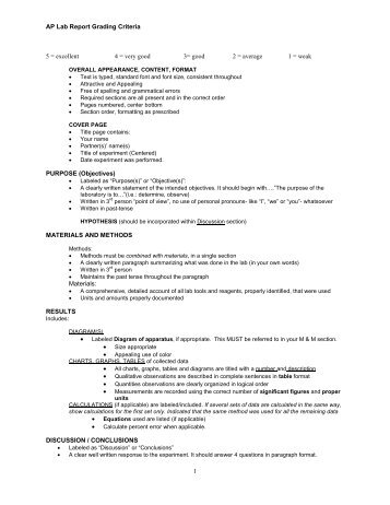
AP Biology Lab 7: Cell Division How do eukaryotic cells divide to produce genetically identical cells or to produce gametes with half the normal DNA? Begin this assignment by reading over this lab in the AP Biology Investigative Lab Manual and reviewing the lab rubric provided in this module. You must complete each section of the lab as directed in the rubric and submit your assignment to the appropriate dropbox. PreLab Assessment Before you begin this lab, review the relevant information in the content and in your textbook. After your review, complete the following prelab assessment.

Lab 11: Animal Behavior. (See The Lab Bench at The Biology. They do not have access to the filter paper and petri dishes recommended in the AP lab manual.

• Click below to review the cell cycle • Click below to review animal mitosis. Cell Division Lab Procedure Materials • pink pipe cleaners • blue pipe cleaners • plastic beads • black marker • Smart Science • Laboratory Investigations Notebook Introduction Review the information provided on pages 83-96 in your lab manual and correlate this information with your prior knowledge of the content to prepare a thorough background, purpose, and hypothesis statement. Be sure to address the concepts of photosynthesis to prepare your background. Use your understanding of scientific methodology to address the purpose of the experiment and prepare a thorough and appropriate hypothesis.
Material & Procedure This investigation consists of five parts, some of which have been altered in order for you to conduct this lab at home. Make sure you understand both the prepared and modified procedure.
Begin by reading over the entire lab very carefully and then create investigative ideas that can be tested by experimentation. Part 1: Modeling Mitosis As indicated on page 87 of your student lab manual, you will use pipe-cleaners to model the phases of mitosis. Follow the directions available on the student worksheet available in the sidebar and record your observations by taking digital pictures of each phase of mitosis. Be sure to answer the questions for Part 1 in your laboratory investigation notebook. Part 2: Effects of Environment on Mitosis Begin by reading all of Part 2 found on pages 87-89 of your student lab manual. As indicated, prepare an experimental and null hypothesis.
Record your hypotheses and the answers to the questions on page 87 in your laboratory investigation notebook. To complete this part of the lab, you will complete a modified version by using Smart Science. Log in using the username and password provided by your teacher and select the Mitosis Lab under the category of Molecules and Cells. Complete the Warm Up Quiz and review the procedure for the lab. Pick Onion Tip 1, watch the video, and select Measure to begin data collection. You will select cells available in the slide that correspond to the different phases of mitosis.
Record the numbers for each phase in your laboratory investigation notebook. Repeat counts using Onion Tip 2 and 3. When you have collected all data, make the necessary calculations and complete Table 1 as indicated on page 88 of your student lab manual. This data will be used as your control. Assume that you also observed cells that have been treated with lectin, how would you expect the numbers to compare to the data collected from your control?
Using your control data and your understanding of the protein lectin, prepare hypothetical data to complete the data table provided below. After you have completed the data table, follow the directions on pg 89 of your student lab manual to calculate the chi-square for your data and answer the post lab review questions in your laboratory investigation notebook. Parts 3 & 4: Loss of Cell Cycle Control in Cancer & Modeling Meiosis Begin by reading the directions for Part 3 found on pages 90-93 in your student lab manual. Review the videos and links about HeLa cells, Henrietta Lacks, Philadelphia Chromosomes, and Meiosis available in the sidebar. After you review this information, answer the questions for Part 3 & 4 in your laboratory investigations notebook.
Select one question to share and discuss in the appropriate discussion forum. Part 5: Modeling Meiosis Read pages 93-96 in your student lab manual. To complete this part of the lab, you will complete a modified version by using Smart Science. Log in using the username and password provided by your teacher and select the Crossing Over in Sordaria Lab under the category of Molecules and Cells. Complete the Warm Up Quiz and review the procedure for the lab.
Pick a wide type and tan (W-T) slide, watch the video, and select Measure to begin data collection. You will count the number of asci that have a 4:4, 2:2:2:2, and 2:4:2 pattern. If you do not have a highly visible slide you can select another slide with a better view to collect your data.
Record the numbers for each pattern from two slides in your laboratory investigation notebook. After you have collected all of your data you can enter the appropriate values in Table 3 as indicated on page 95 of your student lab manual. Remember to record your procedures carefully and in enough detail so that another can easily reconstruct the experiment, however, you do not have to be elaborate. Be careful when constructing your procedure based on the steps provided in the lab manual, as you may have adjusted your procedure to match your needs.
The purpose of this is not to have you recreate the lab that has been prepared, but to use your understanding of scientific methodology and inquiry to construct a viable lab. Data Collection Qualitative data: Using your understanding of the lab, record the qualitative data you collected from the each part of your investigation. Be sure to include pictures from Part 1 in your final lab report. Quantitative data: Organize the data collected from Parts 2 & 5 of this investigation as indicated in your student lab manual. Be sure to label your data tables appropriately.
Analysis & Discussion • Create a graph comparing the control and treated cells from Part 2. • Calculate the chi-square value data collected in Part 2.
Show all work. Indicate if this value results in acceptance or rejection of your null hypothesis. • Include responses to the following questions in your analysis: • What is the significance of the fact that chromosomes condense before they are moved?
• What would happen if sister chromatids failed to separate? • Does an increased number of cells in mitosis mean that these cells are dividing faster than cells in the roots with a lower number of cells in mitosis? • How is the cell cycle controlled in normal cells?
• What are cyclin and cyclin-dependent kinases? • How are normal cells and cancer cells different from each other?
• How was the HeLa cell line cultured? • What are some legal and ethical questions related to the use of HeLa cells? • How do cells monitor DNA integrity? Powershell Form Designer Freeware Dvd on this page.
• The published map distance between the spore color gene and the centromere in Sordaria is 26 map units. How did your data compare? Conclusions Summarize your findings and analysis in complete and coherent paragraphs. Be sure to include the purpose of your experiment and relate your findings to your hypothesis. Also discuss limitations, possible sources of error, and means of improvement for your investigations.
References Include all references in APA format. Don't forget that you should have in-text citations to correlate to the references included in this section. Questions for Thought: HeLa cells, Henrietta Lacks, Philadelphia Chromosomes Part of your responsibility as a scientist is to share your ideas and discoveries with the rest of the scientific community. For AP Lab 7: Cell Division, you were introduced to HeLa cells, Henrietta Lacks, and Philadelphia Chromosomes. Review the videos and links about this information and answer the questions for Part 3 & 4 from your student laboratory manual. Select one question to share and discuss with the class.
Remember, science review uses APA formatting for citation and referencing. Once you've completed your responses, follow your teacher's instructions for submitting your work.
Hand Outs More Resources . Sony Net Md Walkman Mz-n510 Software on this page.
AP Biology Lab 9: Restriction Enzyme Analysis How can we use genetic information to identify and profile individuals? Begin this assignment by reading over this lab in the AP Biology Investigative Lab Manual and reviewing the lab rubric provided in this module. You must complete each section of the lab as directed in the rubric and submit your assignment to the appropriate dropbox. PreLab Assessment Before you begin this lab, review the relevant information in the content and in your textbook. After your review, complete the following prelab assessment.
To prepare for the following lab, it is important for you to have a good understanding of restriction enzyme analysis. Review these concepts by clicking below.
Restriction Enzyme Analysis Procedure Materials Laboratory Investigations Notebook Introduction Review the information provided on pages 111-124 in your lab manual and correlate this information with your prior knowledge of the content to prepare a thorough background, purpose, and hypothesis statement. Be sure to address the concepts of photosynthesis to prepare your background. Use your understanding of scientific methodology to address the purpose of the experiment and prepare a thorough and appropriate hypothesis. Material & Procedure This investigation consists of several activities, some of which have been altered in order for you to conduct this lab at home.
Make sure you understand both the prepared and modified procedure. Begin by reading over the entier lab very carefully and then create investigative ideas that can be tested by experimentation. Activity I: Restriction Enzymes As indicated on page 113 of your student lab manual, you will review the function of restriction enzymes by completing questions 1 & 2 in your student lab manual. Do Not Write in the Lab Manual. Record all observations and responses in your laboratory investigation notebook. Activity II: DNA Mapping Using Restriction Enzymes Read the information for this activity found on pages 114-115 of your student lab manual.
Follow the directions as provided and answer all questions in your laboratory investigation notebook. Activity III: Basic Principles of Gel Electrophoresis Read all the information on pages 115-122. To complete this part of the lab, you will complete a modified version by using a virtual lab from the Genetic Science Learning Center.
Begin by completing the tutorial review provided as part of the virtual lab. After you complete the tutorial, you will be guided to conduct the steps necessary to create and run an electrophoresis gel. Although these procedures are the same as the directions provided on pages 116-119 of your student manual, make sure you review both the lab and manual carefully to ensure thorough understanding. To complete your analysis, read the directions beginning on page 120 of your student lab manual. As indicated, examine the ideal digest provided on page 121 and record the distance traveled for each band from the BamHI, EcoRI, and HindIII digest. Be sure to use a centimeter ruler to record the distance each band from the horizontal and vertical center of the well to the center of the band.
You can print out a copy from the sidebar. Record this data in your copy of Table 1 (DO NOT WRITE IN THE AP LAB MANUAL). As indicated in step 3, you will need to plot the migration distance and the base pair fragment sizes on semilog graph paper from the HindIII digest.
You must use a spreadsheet program, set to graph on a logarithmic scale, otherwise your data will be incorrect. Finally, use the standard curve to calculate the approximate sizes of the EcoRI and BamHI fragments. Record your distance migrated and interpolated sizes (actual bp will be provided when your assignment is graded). To complete this investigation, you will design an investigative technique as discussed in the lab manual and complete the directions as indicated on page 122 of your student lab manual. Remember to record your procedures carefully and in enough detail so that another can easily reconstruct the experiment, however, you do not have to be elaborate. Be careful when constructing your procedure based on the steps provided in the lab manual, as you may have adjusted your procedure to match your needs. The purpose of this is not to have you recreate the lab that has been prepared, but to use your understanding of scientific methodology and inquiry to construct a viable lab.
Data Collection Qualitative data: Using your understanding of the lab, record the qualitative data you collected from the each part of your investigation. For your designed investigation, create an image of a hypothetical gel related to the scenario that will provide proposed quantitative data for analysis. Quantitative data: Organize the data collected from the activities as indicated in your student lab manual and prepare hypothetical data as indicated in your designed investigation. Be sure to label your data tables appropriately. Analysis & Discussion • Create a graph indicating the standard curve created by the HindIII digest. On your graph, indicate the interpolated points of the EcoRI and BamHI digest. • Include the summary of the scenario based on motive, means, opportunity, and DNA evidence.
Include a discussion of your conclusions in the appropriate discussion forum. • Include responses to the following questions in your analysis: • Two restriction fragments of nearly the same base pair size appear as a single band, even when the sample is run to the very end of the gel. What could be done to resolve the fragments? Why would it work? • What is the source of restriction enzymes? What is their function in nature?
• How can a mutation that alters a recognition site be detected by gel electrophoresis? Conclusions Summarize your findings and analysis in complete and coherent paragraphs. Be sure to include the purpose of your experiment and relate your findings to your hypothesis. Also discuss limitations, possible sources of error, and means of improvement for your investigations. References Include all references in APA format.
Don't forget that you should have in-text citations to correlate to the references included in this section. Questions for Thought: Who Dunnit? For AP Lab 9: Restriction Enzyme Analysis, you were presented with a scenario which involved a missing teacher, blood, and several suspects. After conducting the lab, you should have a good idea of how to determine who is responsible for the crime. In your discussion post, describe the scenario based on motive, means, opportunity, and DNA evidence. Be sure to include data collected from your experiment as justification for your conclusions.
Once you've completed your responses, follow your teacher's instructions for submitting your work. .